Ciona intestinalis early embryonic development
Ciona intestinalis early embryonic development ~ from egg to larva ~ >>

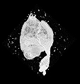
I. Zygote period (0-0.9hr) at 18 degrees C
| Stage | Time after fertilization |
Percentage until hatch |
Tail length (µ m) |
Ratio tail/head |
Description | Link | Image (Normal) |
Image (3D) |
|---|---|---|---|---|---|---|---|---|
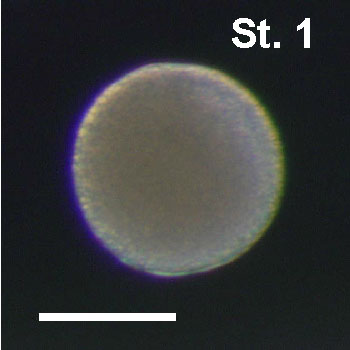
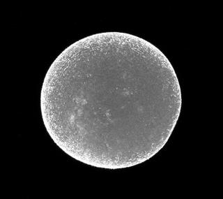
1 |
0.4hr | 2% | - | - | Zygote, fertilized egg |
|
|
|
| 0.8hr | 5% | - | - |
|
|
II. Cleavage period (0.9-4.5hr)
| Stage | Time after fertilization |
Percentage until hatch |
Tail length (µ m) |
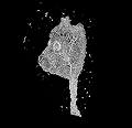
Ratio tail/head |
Description | Link | Image (Normal) |
Image (3D) |
|---|---|---|---|---|---|---|---|---|
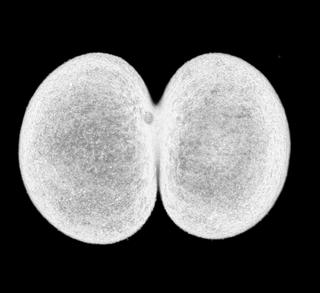
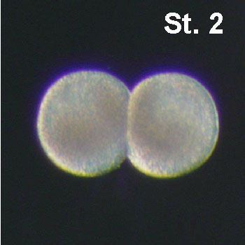
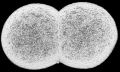
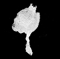
2 |
0.9hr | 6% | - | - | Two cell-stage embryo |
|
|
|
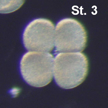
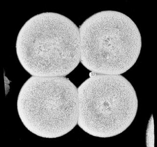
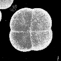
3 |
1.45hr | 8% | - | - | Four cell-stage embryo |
|
|
|
| 1.7hr | 10% | - | - |
|
|
|||
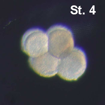
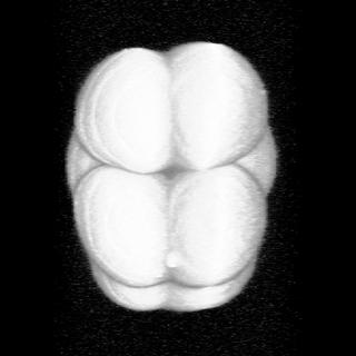
4 |
1.9hr | 11% | - | - | Eight cell-stage embryo |
|
|
|
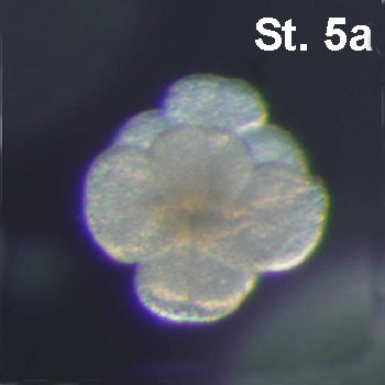
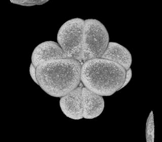
5a |
2.35hr | 13% | - | - | Early sixteen-cell stage embryo |
|
|
|
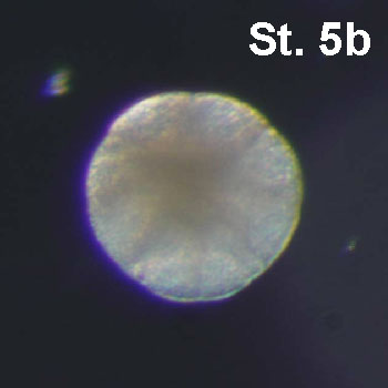
5b |
2.65hr | 15% | - | - | Late sixteen-cell stage embryo |
|
|
|
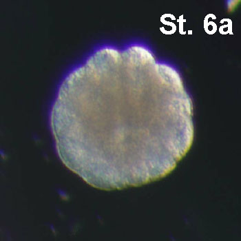
6a |
3hr | 17% | - | - | Early thirty two-cell stage embryo |
|
|
|
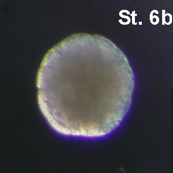
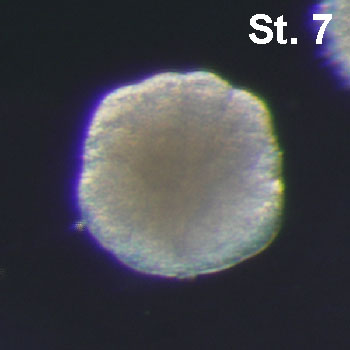
6b |
3.2hr | 18% | - | - | Late thirty two-cell stage embryo |
|
|
|
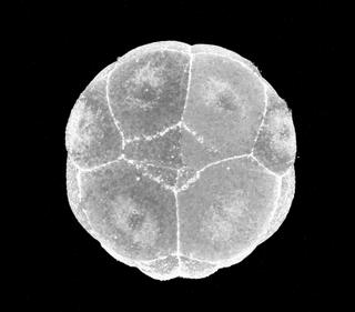
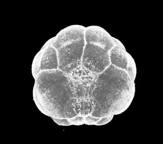
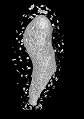
7 |
3.35hr | 19% | - | - | Fourty four-cell stage embryo. The vegetal side of the embryo is very round. |
|
|
|
8 |
4hr | 23% | - | - | Fourty four-cell stage embryo. The vegetal side of the embryo is very round. |
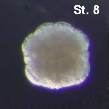
|
|
|
9 |
4.2hr | 24% | - | - | Seventy six cell stage embryo. The vegetal side of the embryo is very flat |
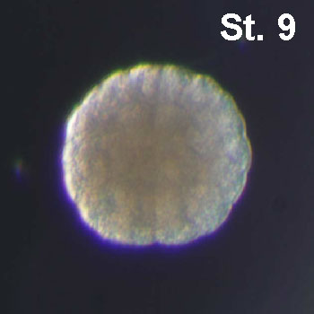
|
|
III. Gastrula Period (4.5-6.3hr)
| Stage | Time after fertilization |
Percentage until hatch |
Tail length (µ m) |
Ratio tail/head |
Description | Link | Image (Normal) |
Image (3D) |
|---|---|---|---|---|---|---|---|---|
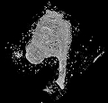
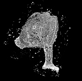
10 |
4.5hr | 26% | - | - | Gastrulation starts with the apical constriction of A7.1 blastomeres. |

|
|
|
| 4.6hr | 27% | - | - |
|
|
|||
11 |
4.9hr | 28% | - | - | The ntochord has invaginated. The vegetal side of the embryo has a horseshoe shape. |
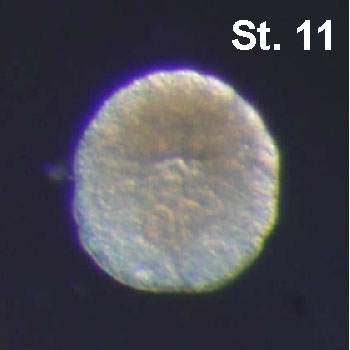
|
|
|
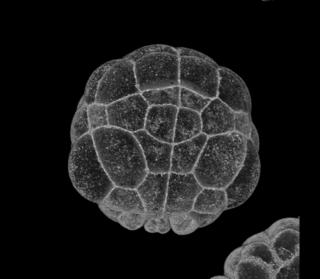
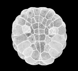
12 |
5.65hr | 32% | - | - | Six-row neural plate stage. The blastopore is still central and open. |
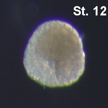
|
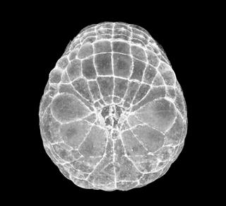
|
|
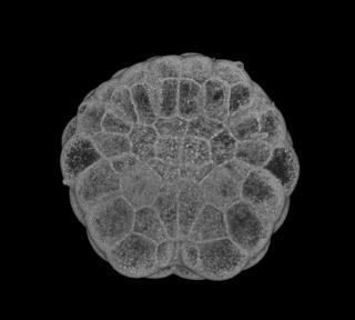
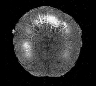
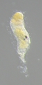
13 |
5.9hr | 34% | - | - | The blastopore is in posterior position and nearly closed. The embryo elongates anteriorly. The neural plate has more than 6 rows and the A-line neural rows (I and II) start to curve (neurulation begin). The large b6.5 progeny are coming together at the midline. |
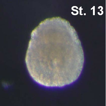
|
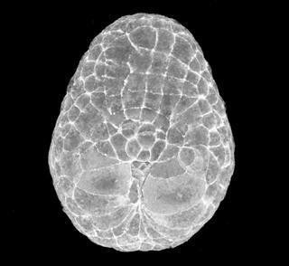
|
IV. Neurula Period (6.3-8.5hr)
| Stage | Time after fertilization |
Percentage until hatch |
Tail length (µ m) |
Ratio tail/head |
Description | Link | Image (Normal) |
Image (3D) |
|---|---|---|---|---|---|---|---|---|
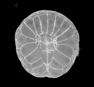
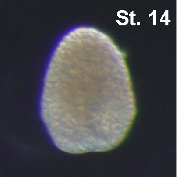
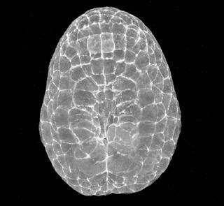
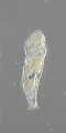
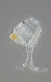
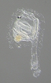
14 |
6.35hr | 36% | - | - | A-line neural plate forms a gutter lined by b6.5 descendants. The embryo has a diamond shape. The gutter is not closed. |
|
|
|
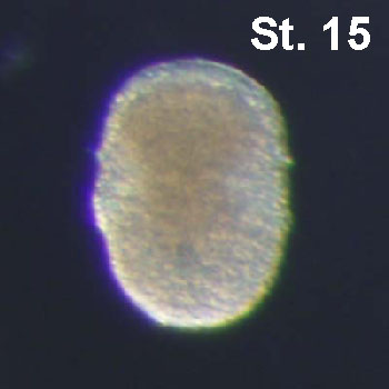
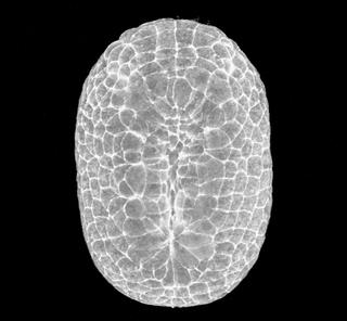
15 |
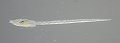
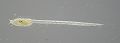
6.8hr | 39% | 83.0 | 1.0 | The neural tube has formed on most of its length. The embryo has an oval shape. The a-line neural plate also forms a gutter. |
|
|
|
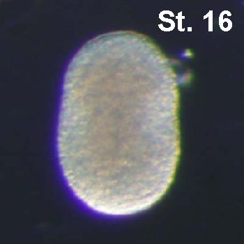
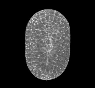
16 |
7.4hr | 42% | 84.2 | 1.0 | The neural tube starts to form in the posterior territories. The embryo elongates. |
|
|
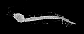
V. Tailbud Period (8.5-17.5hr)
| Stage | Time after fertilization |
Percentage until hatch |
Tail length (µ m) |
Ratio tail/head |
Description | Link | Image (Normal) |
Image (3D) |
|---|---|---|---|---|---|---|---|---|
|
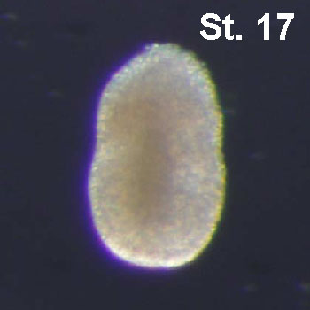
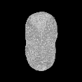
17 |
8.45hr | 48% | 87.8 | 1.0 | First indication of a separation between tail and trunk territories. The tail is not bent and has the same length as the trunk. Any notochord cells not finished intercalation. |
|
|
|
|
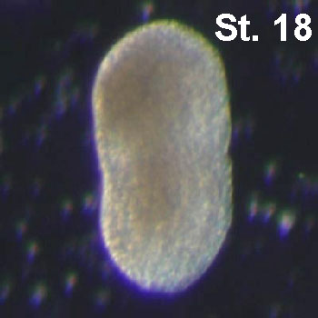
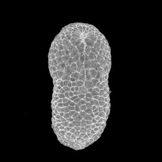
18 |
8.8hr | 50% | 111.9 | 1.1 | The tail is clearly separated from the trunk. Tail and trunk have same length. Neuropore still open, a-line neurulation. |
|
|
|
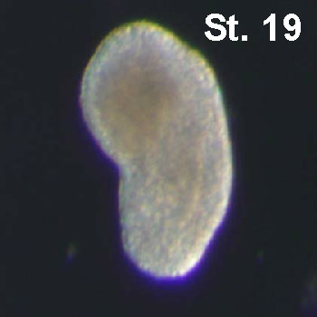
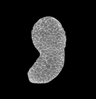
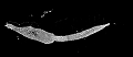
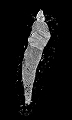
19 |
9.3hr | 53% | 120.3 | 1.2 | The tail bends about 40°and is slightly longer than the trunk. A few anterior most notochord cells begin to intercalate and linear. |
|
|
|
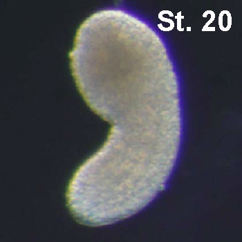
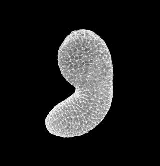
20 |
9.5hr | 54% | 146.7 | 1.3 | Neuropore closed, tail bent by 60°, neurulation complete. |
|
|
|
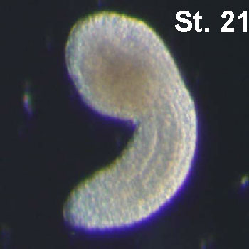
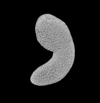
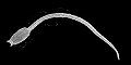
21 |
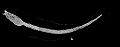
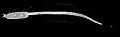
10.0hr | 57% | 180.7 | 1.6 | Tail 1 1/2 times longer than trunk and curve ventrally (90°). Intercalation of notochord cells just finished. |
|
|
|
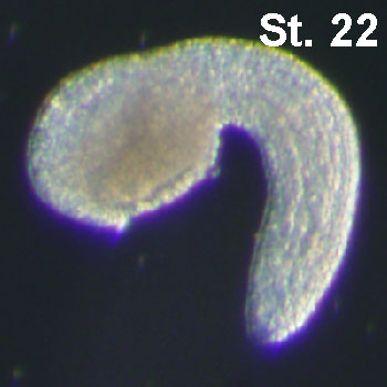
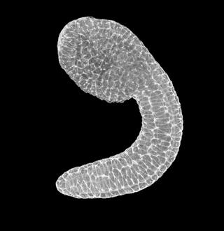
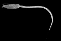
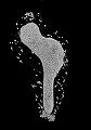
22 |
10.9hr | 62% | 221.2 | 1.9 | The body adopts a half circle shape. Tail twice as long as trunk. |
|
|
|
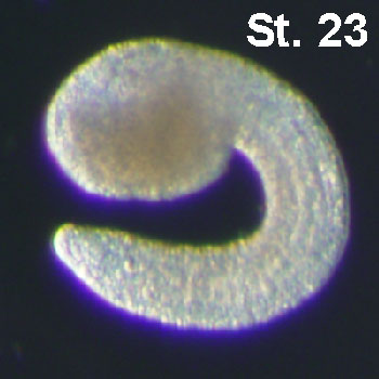
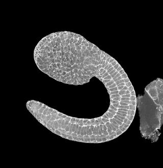
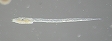
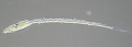
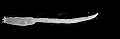
23 |
11.9hr | 68% | 255.1 | 2.1 | Initiation of the pigmentation of the otolith. Tail strongly curved with tip close to the anterior end of the trunk. |
|
|
|
| 12hr | 69% | - | - |
|
|
|||
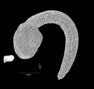
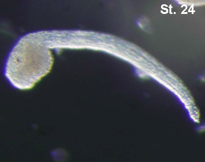
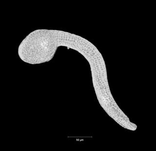
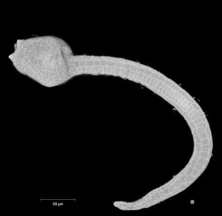
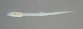
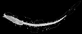
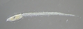
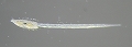
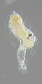
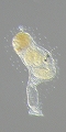
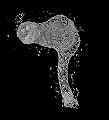
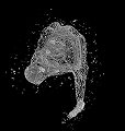
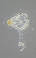
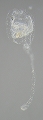
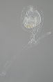
24 |
13.5hr | 77% | 442.6 | 3.4 | Notochord vacuolation begins, palps start to be visible at the front end of the embryo. Tail straightens. |
|
|
|
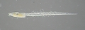
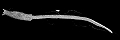
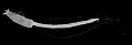
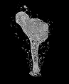
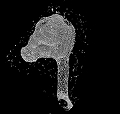
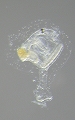
25 |
15.9hr | 91% | 558.6 | 3.9 | Ocellus melanization. All notochord cells have vacuoles. Tail bent dorsally. |
|
|
|
| 16.5hr | 94% | - | - |
|
|
|||
| 17.0hr | 97% | - | - |
|
|
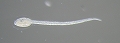
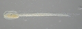
VI. Larva Period (St.26~30, 17.5~23hpf)
*Note that the duration of larval swimming to adhesion differs among individuals. So the time after fertilization during Larva Period was broad.
| Stage | Time after fertilization (18oC) | Description | Link | Image (Normal) |
Image (3D) |
|---|---|---|---|---|---|
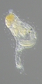
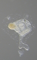
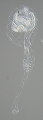
| 17hr 30min (17.5hpf) | Hatching, spherical trunk shape, immature papillae with pyramidal shape, irregular tail movements |
 |
 |
||
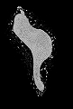
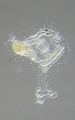
| 17.5-20 hpf | Spindle-like trunk shape, regular tail movements and swimming behaviour |
 |
 |
||
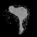
| 20-22hpf | Elongated papillae and expansion of their basal part, square head, spherical test cells, cilia in epidermal sensory neurons recognizable, preoral lobe recognizable |
 |
 |
||
 |
 |
||||
| 22-24hpf | Longer and narrower head with respect to St. 28, trunk profile squared at transition between trunk and tail |
 |
 |
||
 |
 |
VII. Adhesion period (St.31~32, 24~26hpf)
| Stage | Time after fertilization (18oC) |
Description | Link | Image (Normal) |
Image (3D) |
|---|---|---|---|---|---|
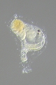
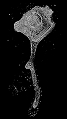
| 24 hpf | Curved papillae, otolith and ocellus remnants recognizable |
 |
 |
||
| 25-27 hpf |
 |
 |
|||
 |
 |
VIII.Tail absorption period (St.31~33, 27~30hpf)
*Matsunobu et al. (2015) showed that the hatched larva requires at least three or four hours to get competence to commence metamorphosis.
| Stage | Time after fertilization (18oC) |
Description | Link | Image (Normal) |
Image (3D) |
|---|---|---|---|---|---|
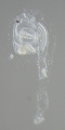
| 27 hpf | Beginnng of tail absorption, tail bending at the transition between trunk and tail, otolith and ocellus remnants recognizable |
|
|
||
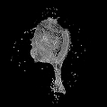
| 28 hpf | 50% of tail absorbed into trunk. Tail shrinked and thickened, otolith and ocellus remnants recognizable |

|
|
||
| 29 hpf | Tail completely absorbed, papiliae no more recognizable, otolith and ocellus remnants recognizable |
|
|
IX. Body axis rotation period (St.34~36, 30~60hpf)
| Stage | Time after fertilization (18oC) |
Description | Link | Image (Normal) |
Image (3D) |
|---|---|---|---|---|---|
| 30-36 hpf | Beginning of body axis rotation (angle between the stalk and the endostyle more than 0°), outer tunic compartment and outer cuticle layer no more present, tunic cells recognizable in definitive tunic, otolith and ocellus remnants recognizable |
 |
 |
||
 |
 |
||||
 |
 |
||||
| 36-45 hpf | Body axis rotation of 30°- 60°, one pair of gill-slit recognizable, otolith and ocellus remnants recognizable |
 |
 |
||
 |
 |
||||
 |
 |
||||
| 45-60 hpf (2dpf) | Two pair of gill-slit open, body axis rotation at 80°-90°, filtering and feeding activity present, otolith and ocellus remnants recognizable, heart beating |
 |
 |
||
 |
 |
||||
 |
 |
||||
 |
 |
||||
 |
 |
X. Juvenile period (St.37~39, 60~168hpf)
| Stage | Time after fertilization (18oC) |
Description | Link | Image (Normal) |
Image (3D) |
|---|---|---|---|---|---|
| 63-72 hpf (3dpf) | Body axis rotation completed, stomach swollen, otolith and ocellus remnants recognizable |
 |
 |
||
 |
 |
||||
 |
 |
||||
 |
 |
||||
38 early juvenile II |
4 dpf | Larval tail remnants totally adsorbed |
 |
 |
|
 |
 |
||||
39 mid juvenile I |
6 dpf | Additional gill slit begin to open, appearance of stomach, gut and neural grand |
 |
 |
|
 |
 |