Developmental table
I. Zygote period
Stage
Image
3D
Slice
DIC
hpf
Simple description
Corresponding Stages in Ciona
1
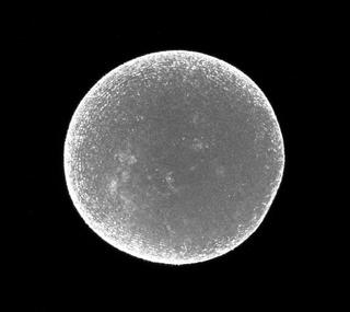
Fertilized egg
Fertilized egg


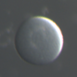
0 hpf
Zygote, Fertilized egg


-
II. Cleavage period
Stage
Image
3D
Slice
DIC
hpf
Simple description
Corresponding Stages in Ciona
2
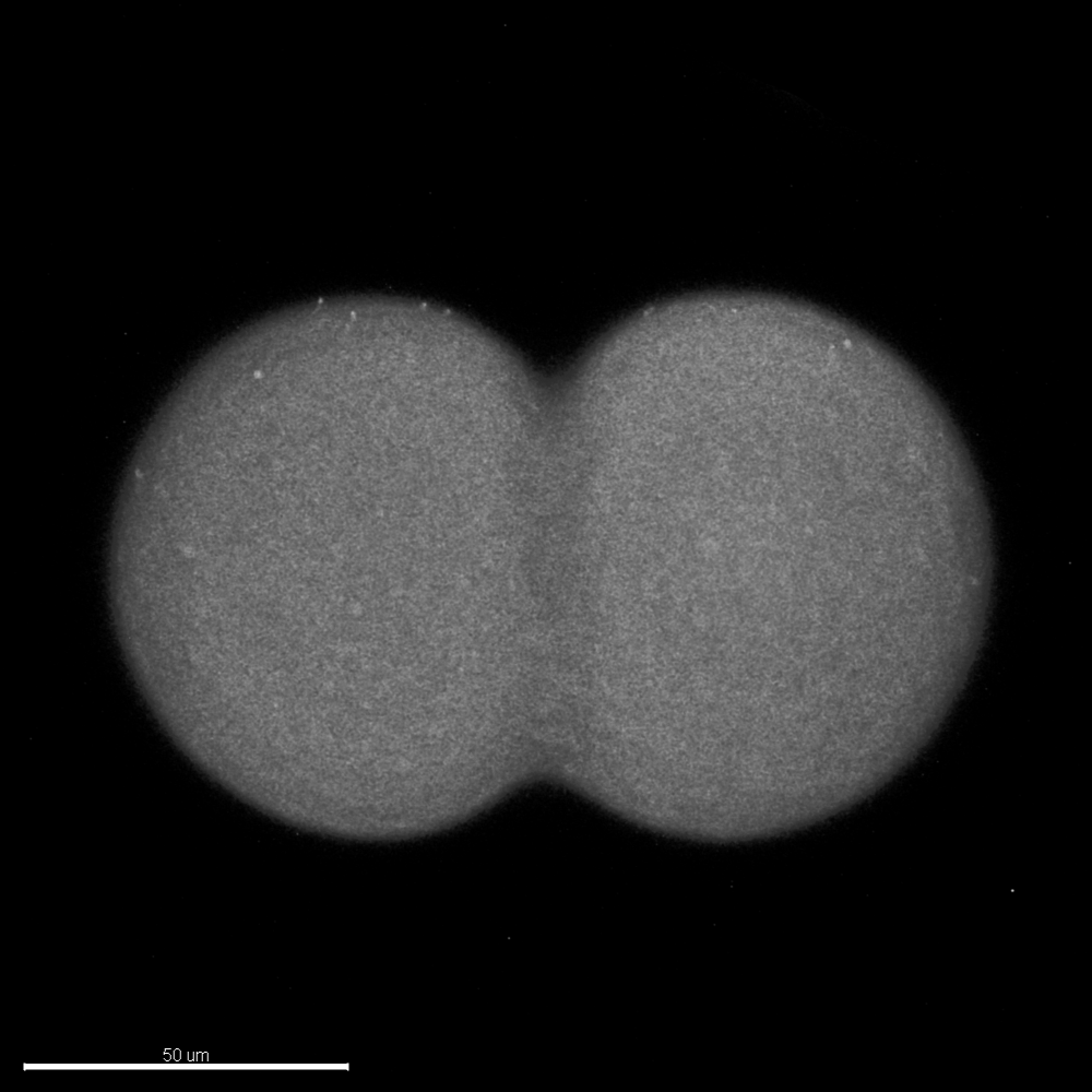
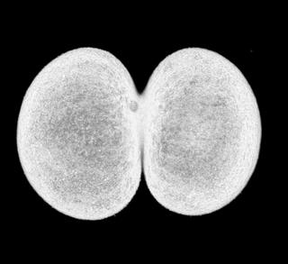
Two-cell
Two-cell


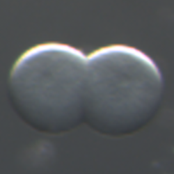
1.2 hpf
The embryo composed of two cells.

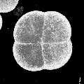
3
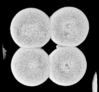
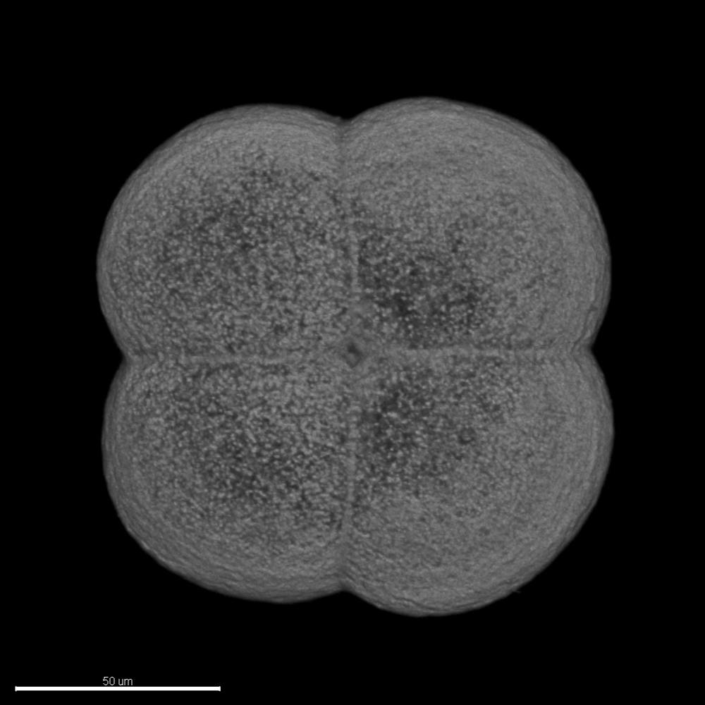
Four-cell
Four-cell


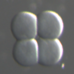
1.5 hpf
The embryo composed of four cells.


-
4
Eight-cell
Eight-cell


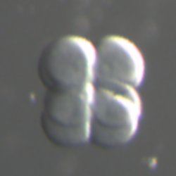
1.9 hpf
The embryo composed of eight cells.
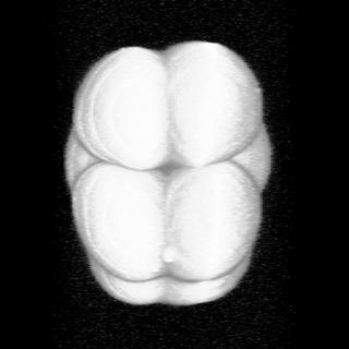
5a
Early 16-cell
Early 16-cell
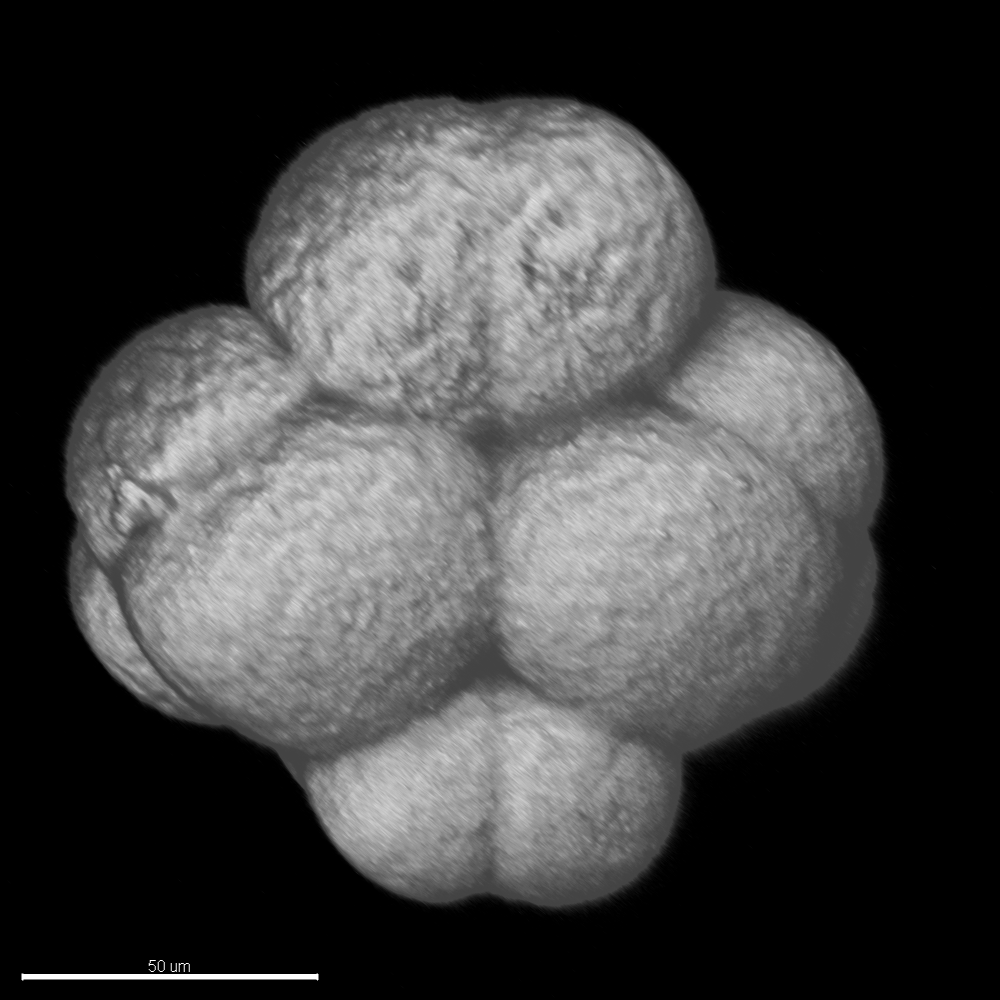

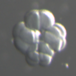
2.3 hpf
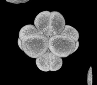
The embryo composed of 16 cells. Blastomeres is
uncompacted in this stage.
5b
Late 16-cell
Late 16-cell
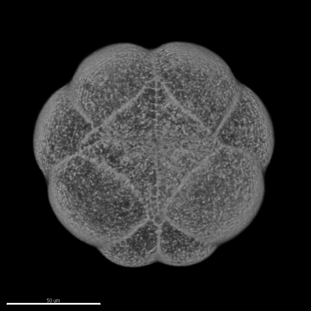

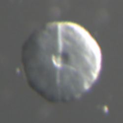
2.5 hpf
The embryo composed of 16 cells. Blastomeres has been
compacted.
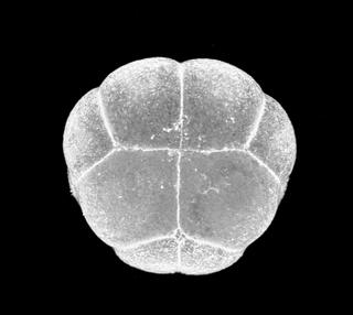
6a
Early 32-cell
Early 32-cell
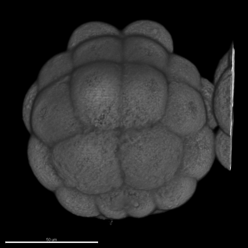

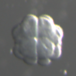
2.8 hpf
The embryo composed of 32 cells. Blastomeres is uncompacted in
this stage.
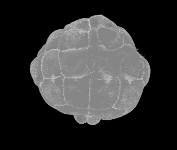
6b
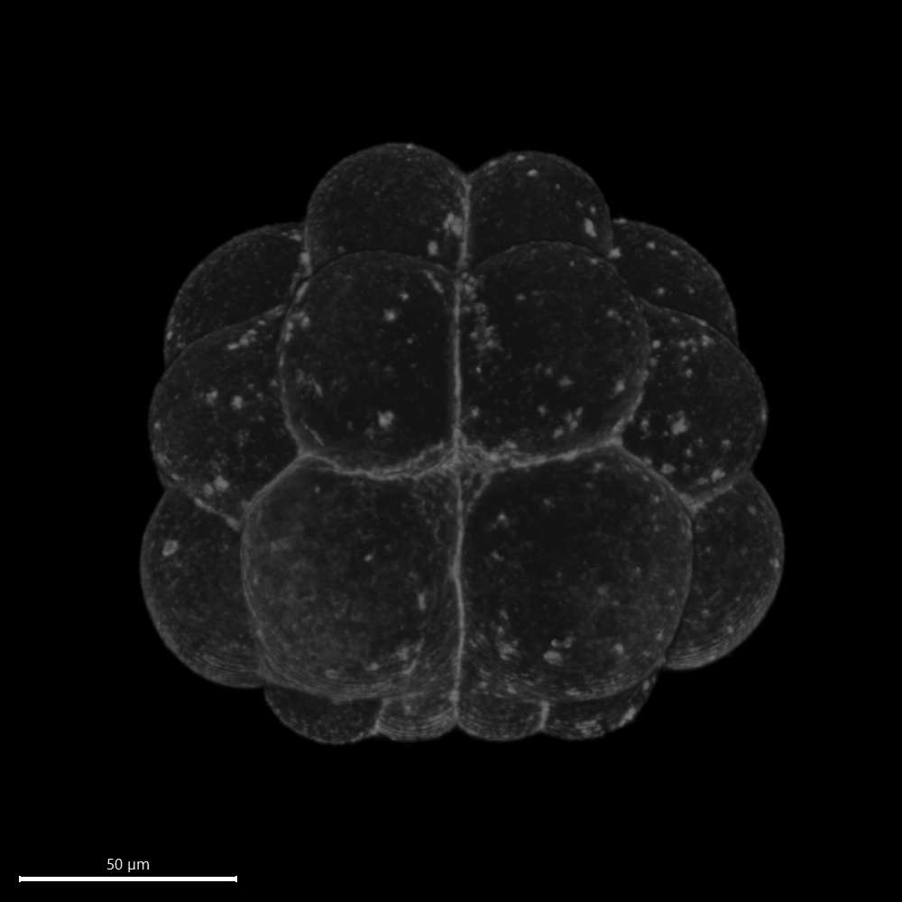
Late 32-cell
Late 32-cell

3.0 hpf
The embryo composed of 32 cells. Blastomeres has been
compacted.
7
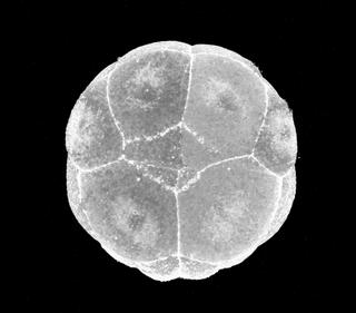
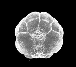
44-cell
44-cell


3.3 hpf
The embryo composed of 44 cells. The vegetal side blastomeres
are bulging out.
8
64-cell
64-cell


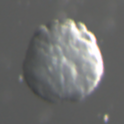
3.6 hpf
The embryo composed of 64 cells. Embryo has an almost circle
shape from top view.
9
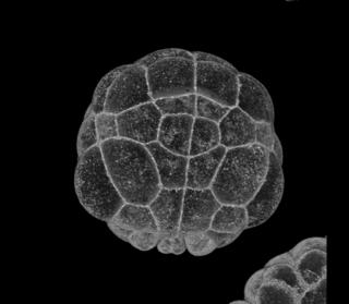
76-cell
76-cell


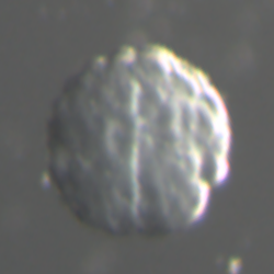
4.0 hpf
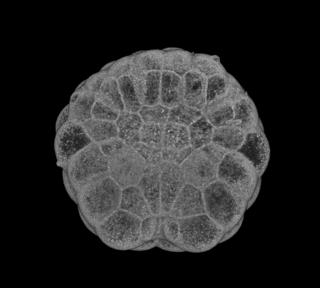
The embryo composed of 76 cells. The embryo flattens on its
vegetal side,
in preparation for gastrulation.
III. Gastrula period
Stage
Image
3D
Slice
DIC
hpf
Simple description
Corresponding Stages in Ciona
10
Initial gastrula,
112-cell
Initial gastrula,
112-cell


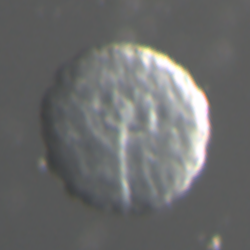
4.4 hpf
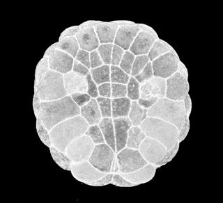
Gastrulation starts with A7.1 blastomeres, which is the center
of invagination.
11
Early gastrula
Early gastrula


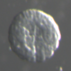
4.8 hpf
The notochord has invaginated. The vegetal side of the embryo
has a horseshoe shape.
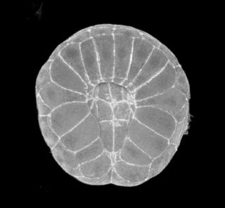
12
Mid gastrula
Mid gastrula


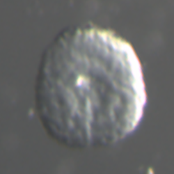
5.3 hpf
The embryo has six-row neural pLate. The blastopore is located
posterior and still open.
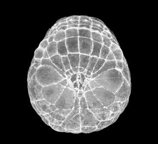
13
Late gastrula
Late gastrula


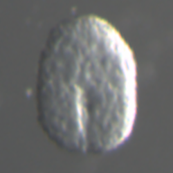
5.7 hpf
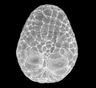
The embryo elongates anteriorly. The blastopore is located
posterior and nearly closed. The neural plate has more than 6 rows and a part of neural rows
start to curve.
IV. Neurula period
Stage
Image
3D
Slice
DIC
hpf
Simple description
Corresponding Stages in Ciona
14
Early neurula
Early neurula


6.2 hpf
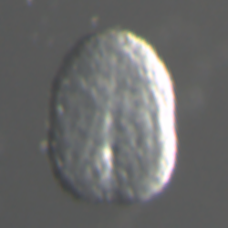
Neural plate forms a furrow. The embryo has an oval shape. The
furrow is not closed.
15
Mid neurula
Mid neurula


6.7 hpf
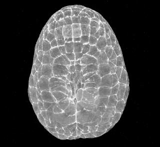
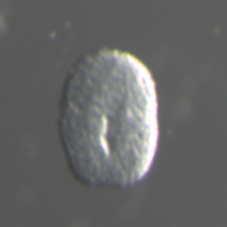
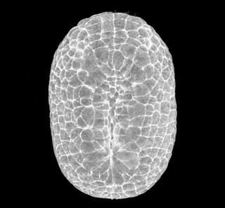
The neural tube has formed along most of its length. The
embryo has an oval shape. The A-line neural plate also forms a neural fold.
16
Late neurula
Late neurula


7.3 hpf
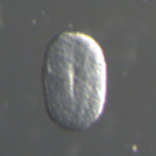
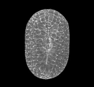
The neural tube closure starts in the posterior part. The
embryo more elongates along A-P axis.
V. Tailbud period
Stage
Image
3D
Slice
DIC
hpf
Simple description
Corresponding Stages in Ciona
17
Initial tailbud I
Initial tailbud I


8.0 hpf
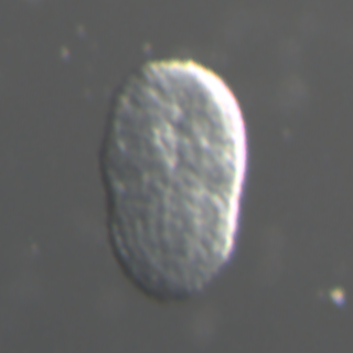
First indication of a separation between trunk and tail parts
in this stage. The tail is not bent and boundary area between trunk and tail parts bulging
out. The neural tube closure in the posterior territory finished and the neuropore move more
anterior. Any notochord cells not finished intercalation.
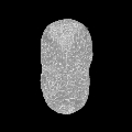
18
Initial tailbud II
Initial tailbud II


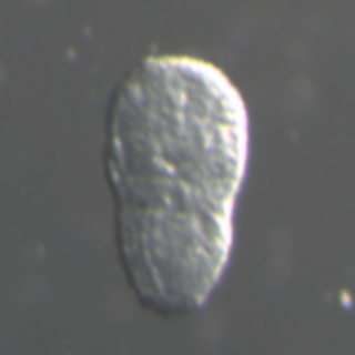
8.3 hpf
The tail is clearly distinguished from the trunk and begins to
bend. The tail is shorter than the trunk. The neuropore is still opened.
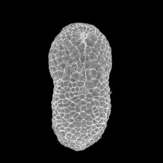
19
Early tailbud I
Early tailbud I


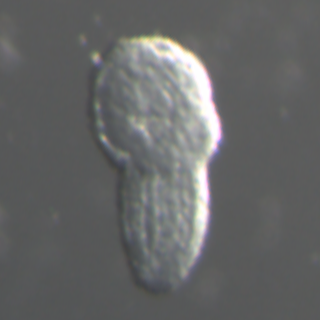
8.8 hpf
The angle formed by trunk and tail is an obtuse angle and has
the same length as the trunk. A few anterior notochord cells finish intercalation and the
neuropore just close.
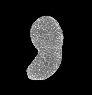
20
Early tailbud II
Early tailbud II


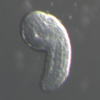
9.2 hpf
The tail bends about 90° and a half of notochord cells finish
intercalation. The neuropore has closed.
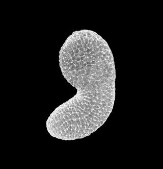
21
Mid tailbud I
Mid tailbud I


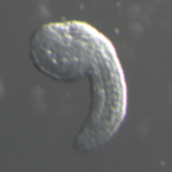
9.7 hpf
The tail is 1 1/2 times longer than the trunk and the angle
formed by trunk and tail is an acute angle. Intercalation of the notochord cells is
completed.
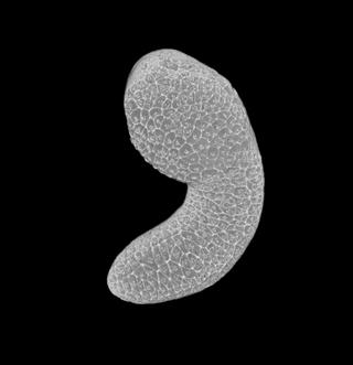
22
Mid tailbud II
Mid tailbud II


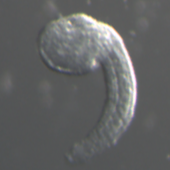
10.1 hpf
The body curves circularly.
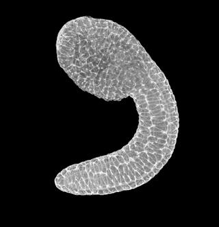
23
Late tailbud I
Late tailbud I


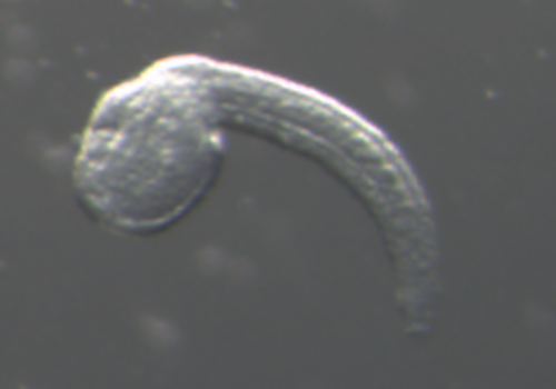
10.5 hpf
The pigmentation of the otolith starts. The tail most strongly
curved in this stage and the tail length is twice as long as trunk.
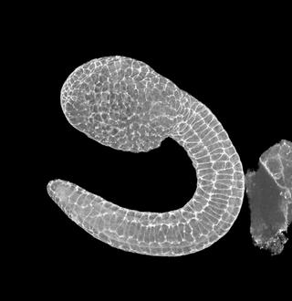
24
Late tailbud II
Late tailbud II


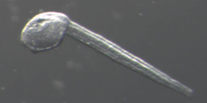
12.1 hpf
The notochord vacuolation begins partially and palps formation
is initiated by the anterior trunk epidermis thickening and bulging. Tail straightens in its
anterior part.
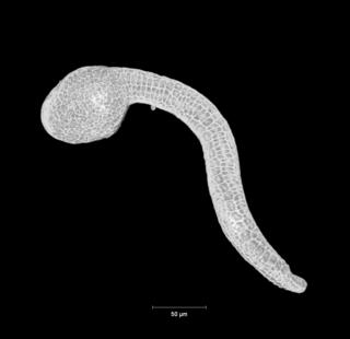
25
Late tailbud III
Late tailbud III


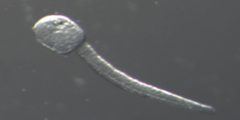
13.3 hpf
The ocellus melanization is observed. The cilia elongation
from caudal epidermal neuron begins. All notochord cells have vacuoles.
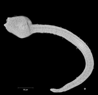
VI. Larva period
Stage
Image
3D
Slice
DIC
hpf
Simple description
Corresponding Stages in Ciona
26
Hatching larva
Hatching larva


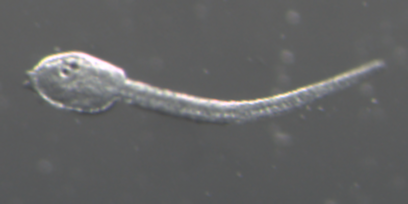
16.2 hpf
Larvae are hatching. The trunk has an elongated rectangular
shape.