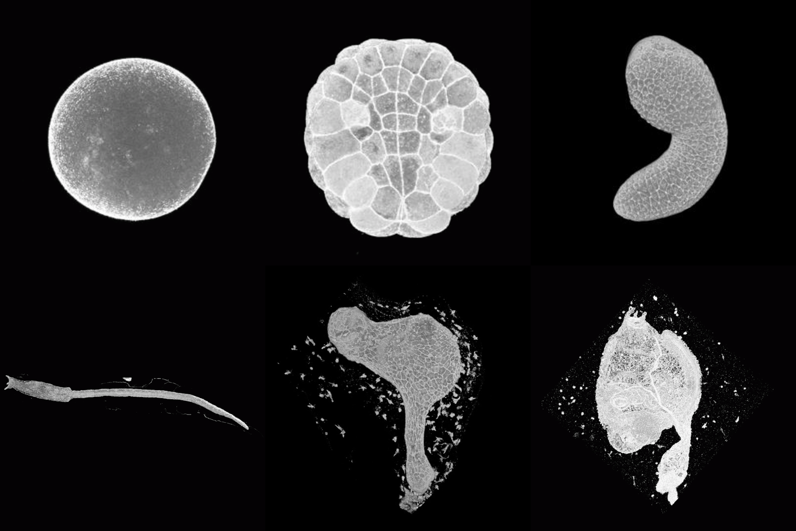
In solitary ascidian, Ciona intestinalis type A (Ciona robusta) *, there occur important developmental events after hatch, such as larval tissue maturation of nervous system and tail tissues for swimming motility and subsequent a series of metamorphosis to become adult. The ascidian metamorphosis divided into following 5 different sub-processes, adhesion to the ground by papilla, apoptotic degeneration of tail and absorption of larval tissue, removal of larval tunic, rotation of body axis and differentiation of adult organs. So the description of the ascidian development after hatching larva is important to understand the apoptosis of larval organ and the origin of adult organ. However, there is no spatio-temporal description at cellular level in this period.
Previously, we made the standard web-based image resource called Four-dimensional Ascidian Body Atlas, "FABA", including ascidian's three-dimensional (3D) and cross-sectional images through the developmental time course. These images were acquired by visualizing F-actin of ascidian, C. intestinalis, stained with alexa fluor phalloidin by confocal laser scanning microscope (CLSM) and reconstructed from more than 3,000 high-resolution real images at newly defined 26 distinct developmental stages from fertilized egg to hatching larva (Hotta et al., 2007, https://www.bpni.bio.keio.ac.jp/chordate/faba/1.4/top.html).
In this study, to clear spatio-temporal morphological change of cells after hatching and construct the comprehensive anatomical and developmental ontology, "TUNICANATO", we applied above mentioned approach to Ciona in late developmental stages, from 17.5hpf hatching larva to 7dpf juvenile. So far, time-lapse images by stereo microscope were performed and matched with images by CLSM which correspond to 31 different developmental timings. By using these dataset, we could trace ascidian development at single cell/tissue level until juvenile. TUNICANATO will be a standard database for ontology of development and anatomy in Ciona.
Our dataset will be helpful in standardizing developmental stages for morphology comparison as well as for providing the guideline for several functional studies of cell biology in adult ascidian.
Please refer to:
